
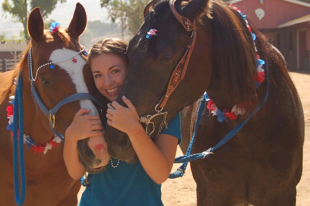
CASE HISTORY
|
California Equine Orthopedics
Mark Martinelli, DVM, DACVS, PhD Lea Walker, DVM www.calequorth.com |
|
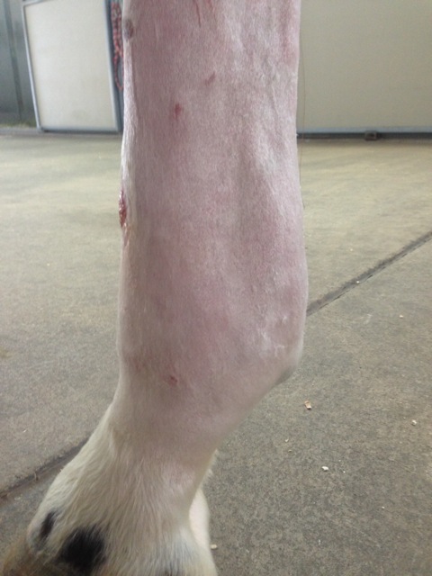
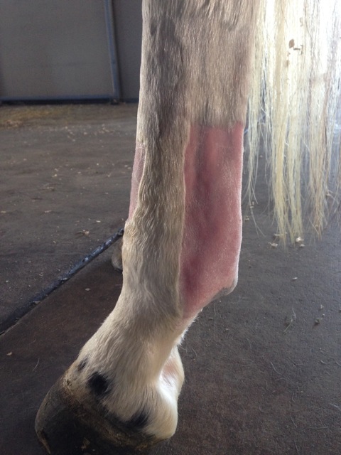
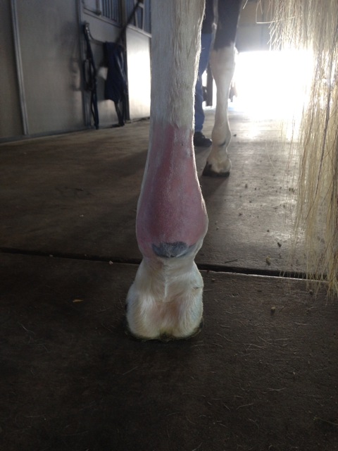
Pre- Tx notes:
Ultrasound Interpretation: In the left hindlimb, there is significant thickening of the annular ligament characterized by fiber separation, fiber disruption and scar tissue accumulation. The palmar surface of the superficial digital flexor tendon also shows significant fiber disruption and separation. Dynamic examination revealed loss of the normal independent gliding motion of the flexor tendons, consistent with an adhesion between the annular ligament and the SDFT. Horse History:
|

|
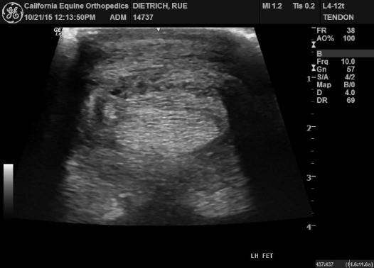
Findings and notes here Conclusion:
Ultrasound Interpretation: In the left hindlimb there is significant reduction in the size of the annular ligament associated with an improved fiber pattern of both the annular ligament and the palmar surface of the SDFT. Dynamic examination revealed significant improvement in the independent gliding motion of the tendons, however evidence of adhesions were still present.

End of RLT Tx: Feb 3, 2016
using Logic E Follow-Up Tool, new images on R
| |||||||
|
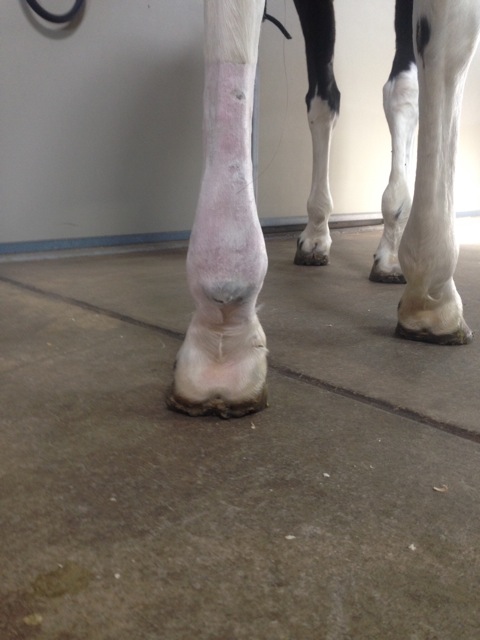
60 Day Recheck:
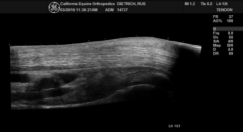
March 30, 2016: Ultrasound Interpretation: In the left hindlimb, the size and fiber pattern of the annular ligament is significantly improved, characterized by no significant regions of fiber separation or disruption. The SDFT is also improved with no significant fiber separation or disruption identified on the palmar surface of the tendon. Dynamic examination revealed return to the normal independent gliding motion of the flexor tendons. 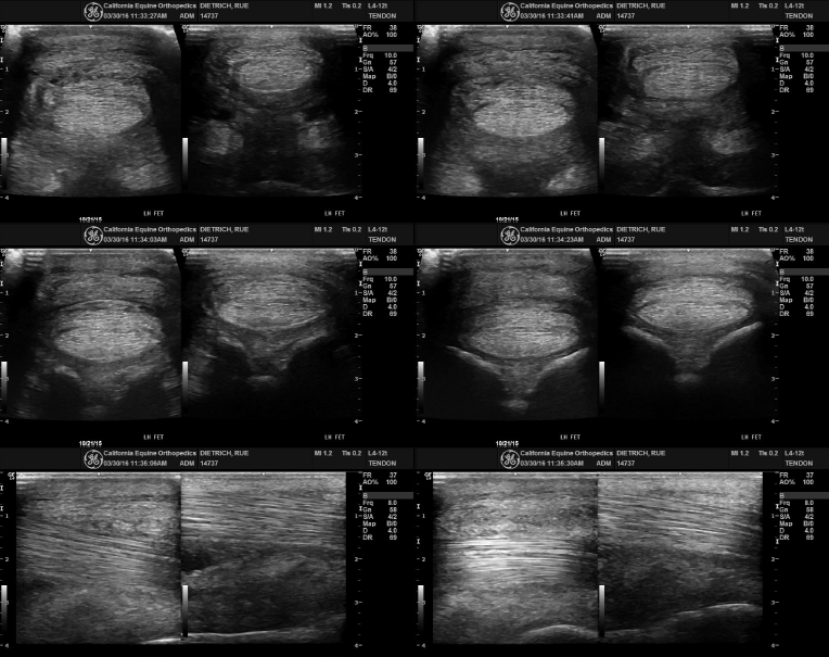
3/30/2016
|
